Nanda - Nursing Care Plan

Pathophysiology and Clinical Manifestations of Diarrhea

Nursing Care Plan for Diarrhea

Diarrhea is an increase in the frequency of bowel movements, an increase in the looseness of stool or both.

Diarrhea is caused by increased secretion of fluid into the intestine, reduced absorption of fluid from the intestine or rapid passage of stool through the intestine.

Pathophysiology of Diarrhea

The basic mechanisms that cause diarrhea are the first osmotic disruption, due to the presence of food or substances that are not absorbed will cause the osmotic pressure within the gut cavity rises, resulting in a shift of water and electrolytes into the intestinal cavity, excessive intestinal cavity contents, will stimulate the intestines to remove, causing diarrhea.

Second, due to certain stimuli (such as toxins) in the gut wall will be an increase of water and electrolytes into the gut cavity and subsequent diarrhea arises because there is an increase in intestinal cavity contents.

Third, motalitas intestinal disorders, the hyperperistaltic, will lead to less opportunity to absorb food intestine causing diarrhea, conversely, when the peristaltic intestinal bacteria decreases will result in excessive arise later can also cause diarrhea.

Besides diarrhea may also occur, due to the entry of microorganisms living in the gut after successfully passing through the stomach acid barrier, microorganisms proliferate, then release toxins and toxin occurs due to hypersecretion which in turn will cause diarrhea.

While the result of the diarrhea will occur of the following:

1. Loss of water (dehydration)

Dehydration occurs due to water loss (output) more than income (input), is the cause of death in diarrhea. Disorders of acid-base balance (metabik acidosis)
It occurs due to loss of Na-bicarbonate with feces. Lipid metabolism is not perfect so dirty stuff accumulate in the body, the accumulation of lactic acid due to anorexia tissue. Acidic metabolic products are increasing due to be issued by the kidneys (oliguria occurs / anuria) and the removal of Na ions from the extracellular fluid into the intracellular fluid.

2. Hypoglycemia

Hypoglycemia occurs in 2-3% of children suffering from diarrhea, more often in children who had previously been suffering from protein-calorie deficiency. This occurs because of the interference of storage / supply of glycogen in the liver and disruption of glucose absorption. Symptoms of hypoglycemia will occur if the blood glucose levels were 40 mg% in infants and 50% in children.

3. The occurrence of weight loss in a short time, this is caused by:
Food is often stopped by parents, for fear of diarrhea or vomiting intensified.
Although milk passed, often given to expenditures and of diluted milk was given too long.
Food given often can not be digested and absorbed properly because of hyperperistaltic.

4. Impaired circulation
As a result of diarrhea, hypovolemic shock can occur, resulting in reduced tissue perfusion and hypoxia, acidosis gain weight, can cause brain bleeding, decreased consciousness, and if not addressed client will die.


Clinical Manifestations of Diarrhea
  1. At first, children / baby whiny anxiety, body temperature may be increased, decreased appetite.
  2. Frequent bowel movements with liquid or watery stool consistency.
  3. Stool color changed to greenish due to mixed bile.
  4. Anus, and surrounding blisters because of frequent bowel movement and stool becomes more acidic due to the amount of lactic acid.
  5. There are signs and symptoms of dehydration, clear skin turgor, fontanel and sunken eyes, dry mucous membranes and with weight loss.
  6. Changes in vital signs, pulse and respiration rapid drop in blood press, rapid heart rate, the patient is very weak, decreased consciousness (apathy, samnolen, sopora komatus).
  7. Reduced diuresis (oliguria to anuria).
  8. If metabolic acidosis occurs, the client will look pale and breathing fast and deep.

7 Nursing Diagnosis for Diabetic Ketoacidosis


Diabetic Ketoacidosis is a medical condition which most likely occurs in patients with Type 1 Diabetes. It occurs when the body cannot use sugar (glucose) as a fuel source because there is no insulin or not enough insulin. Fat is used for fuel instead.

Diabetic Ketoacidosis is a state of absolute or relative insulin deficiency aggravated by ensuing hyperglycemia, dehydration, and acidosis-producing derangements in intermediary metabolism. The most common causes are underlying infection, disruption of insulin treatment, and new onset of diabetes.

Another term for this complication is DKA. Although it is present in Type 1 Diabetes patients, they can also be found in patients who are classified as Type 2 Diabetes. In some instances, this condition is the initial sign that determines people are diabetic.

Symptoms can include:
  • Deep, rapid breathing
  • Nausea and vomiting
  • Dry skin and mouth
  • Stomach pain
  • Flushed face
  • Fruity smelling breath

Other symptoms that can occur include:
  • Breathing difficulty while lying down
  • Abdominal pain
  • Decreased consciousness
  • Decreased appetite
  • Dulled senses that may worsen to a coma
  • Fatigue
  • Muscle stiffness or aches
  • Shortness of breath
  • Frequent urination or thirst that lasts for a day or more
  • Headache

The treatment for Diabetic Ketoacidosis often involves fluid replacement orally or through an IV, electrolyte replacement, and insulin therapy. There are also other instances wherein the patient will need further treatment. When this happens, it is best to follow what the physician recommends.


7 Nursing Diagnosis for Diabetic Ketoacidosis

1. Fluid Volume Deficit
related to:
osmotic diuresis due to hyperglycemia,
excessive discharge: diarrhea, vomiting; restriction intake due to nausea, mental mess.

2. Imbalanced Nutrition: Less Than Body Requirements
related to:
insufficiency of insulin,
decreased oral input,
hipermetabolisme status.

3. Risk for Infection (sepsis)
related to:
increased levels of glucose,
decreased leukocyte function,
changes in the circulation.

4. Risk for Sensory-Perceptual Alterations
related to:
ketidkseimbangan glucose / insulin and / or electrolytes.

5. Fatigue
related to:
decreased metabolic energy production,
insufficiency of insulin,
increasing energy demand: status hypermetabolic / infection.

6. Powerlessness
related to:
long-term illness,
dependence on others.

7. Knowledge Deficit: the disease, prognosis, and treatment
related to:
interpreting the error information,
do not know the source of information.

Acute Pain - Nursing Care Plan for Pneumonia

Pneumonia is an inflammation of the lungs that is usually caused by infection.

The most common causes of pneumonia are infections caused by:
  • bacteria - the most common cause of pneumonia in adults
  • viruses - often responsible for pneumonia in children
  • mycoplasma - organisms that have characteristics of bacteria and viruses that cause milder infections
  • opportunistic organisms - a threat to people with vulnerable immune systems (e.g., Pneumocystis carinii pneumonia in people who have AIDS)

A person who has a higher risk of pneumonia:
  • is under one year of age or over the age of 65
  • is a smoker
  • has a cold or flu
  • has a weak immune system due to cancer therapy, HIV infection, or other disease
  • is undergoing surgery
  • has a problem with alcohol use
  • has a chronic illness such as heart disease, lung disease, or diabetes
  • has a chronic lung disease, such as asthma or chronic obstructive pulmonary disease


Nursing Diagnosis for Pneumonia : Acute Pain related to inflammation of the lung parenchyma, cough settled

characterized by:
  • chest pain
  • headache
  • restless

Goal: pain can be resolved

Expected outcomes:
  • Say no pain / pain can be controlled
  • Shows relax, rest or sleep and increased activity appropriately.

Intervention and Rational:

Independent
  • Evaluation of patient response to activity. Note the report dyspnea. Increased weakness or fatigue, and changes in vital signs during and after activity.
  • Determine the characteristics of the pain, ie sharp, constant, stabbed, explore the changing character / location / intensity of pain
  • Monitor vital signs
  • Provide convenient measures, such as back massage, change of positions, music, quiet / conversation, relaxation / breathing exercises.
  • Advise and assist the patient in mechanical chest compressions during episodes of coughing

Collaboration
  • Give analgesic and antitussive as indicated

Rationale :
  • Setting abilities / needs and facilitate patient choice of intervention.
  • Chest pain, usually within a few degrees of pneumonia, pneumonia can also arise complications such as pericarditis and endocarditis.
  • Changes in heart rate or blood pressure showed that patients experiencing pain, especially when other reasons for changes in vital signs have been seen.
  • Non-analgesic action is given a touch slow to eliminate the discomfort and maximize the effects of analgesic therapy.
  • Tool to control chest discomfort while increasing the effectiveness of cough effort.
  • These drugs can be used to suppress non-productive cough / paroksimal convenience / rest.

Communication Management in Organizations


Communications management is the systematic planning, implementing, monitoring, and revision of all the channels of communication within an organization, and between organizations; it also includes the organization and dissemination of new communication directives connected with an organization, network, or communications technology.

Communication within the organization are: Communication in an organization, which carried out the leadership, both with employees and with an audience that has to do with the organization, in order to develop a harmonious working together to achieve the goals and objectives of the organization.

Organizational communication, can be defined as the performance and interpretation of messages between communication units that are part of a particular organization. An organization consists of units of communication in hierarchical relationships between each other and work in an environment.

Organizational communication occurs whenever at least one person occupying a position in an organization interpret the show. Because the focus is communication among the members of an organization. Organizational communication analysis involves a review of the many transactions that take place simultaneously.

The simplest model of communication is the sender, message and receiver.

If one element is missing, the communication can not take place. For example, one can send a message, but if nothing is received or a hearing, the communication will not occur.

A detailed model of communication, with the essential elements of an organization are:
1. Sources have any ideas, thoughts or impressions
2. translated or encoded into words and symbols, then
3. delivered or sent as a message to the recipient
4. receiver capture symbols and
5. back translated or interpreted back into an idea and
6. send various forms of feedback to the sender.

Guidelines for good communication

1. Researching the real goal in any communication.
2. Consider the physical and psychological state of others in communicating.
3. Consult with various parties every manejemen process from planning to evaluation.
4. Note the pressure tone and facial expressions according to the content of the message.
5. Note the consistency in communication.
6. Be a good listener in communication.

Colds and How to Overcome


Colds flu virus can be said not too dangerous, but it can lower your immune system and make you more susceptible to a more dangerous virus. Despite issuing snot, it seems the best way to get rid of nasty germs, but it turned out to be the body's ability to fight germs.

Unfortunately, issued snot, leading to sinus blockage by mucus full of germs. Research shows that when a person is issued a snot, it can reverse the flow of mucus into the sinuses.

People who issued Snot, can increase pressure in the nasal cavity and push mucus back into the sinuses. The authors state that, way back in the sinus mucus can lead to the development of infection.

Better wipe the snot flow out of the nose. Wiping the slime does not cause mucus back into the sinuses and allow germs out of the nasal cavity naturally, so you can quickly recover from the flu.

Natural way to remove Snot from the sinus is to rinse the nasal cavity with salt water using basin. Add half a teaspoon of sea salt into a cup of warm water, or add antiseptic if the flu is accompanied by a sore throat.

In order to clean the nasal cavity, you can also use a nasal spray containing hydrogen peroxide (certainly should not be many) after gargling with salt. Next, spray into both nostrils, pinch the nose with a finger and tilt the head forward so that the drug reaches the upper sinus cavity. When the fluid flows out of the nose, do not blow, but wiping.

Sample of Nursing Care Plan for Typhoid Fever - 4 Diagnosis and Interventions


Assessment 

Today, Ms.. W age 20, came to the hospital with complaints of high fever (39 0 C) since yesterday. Other complaints: headache, nausea, cough, also complained a few days no bowel movement, the client says insomnia and frequent waking, the client says weak to do an activity, the client complains of pain abdomen, the client says taste in the mouth, and body weakness. After vital sign and physical examination found data: blood pressure: 120/70 mmHg, pulse: 110 x / min, respiration: 30 x / min, typical tongue state (white, dirty, edges hyperemia, chapped lips, face red, lots of sweat). Then the client perform laboratory tests (test widal: 500, check full blood: leukocytes more than 500 / mm3, and SGOT-SGPT) all the results are positive. Client installed oxygenation, the client looks nervous, looking grimacing in pain, weakness, weight decreased, looks bedrest, and assisted activity, faecal mass palpable in the lower abdomen, the portion of food that is provided is not exhausted, vomiting. His family said earlier do not want to be taken to hospital because he felt the disease would heal itself but after the heat higher, clients increasingly anxious and eventually want to be taken to hospital. Also obtained information that the client's father had a history of hypertension.

5 Nursing Diagnosis for Typhoid Fever

Diagnoses that may appear on the client with Typhoid Fever are:

1. Risk for Fluid Volume Deficit related to hyperthermia, a lot of sweat and vomit

characterized by chapped lips, skin turgor ugly.

Goal: fluid volume imbalance does not occur

Expected outcomes:

Mucous membranes moist lips, vital signs (blood pressure, temperature, pulse, respiration) within normal limits, no signs of dehydration.

Intervention:

Assess for signs of dehydration such as dry mouth mucosa, inelastic skin turgor, and increased body temperature, monitor fluid intake and output in 24 hours, measuring body weight each day at a time and at the same time, the report noted such things as nausea, vomiting , stomach pain and distortion. Encourage clients to drink plenty of approximately 2000-2500 cc per day, collaboration in laboratory tests (Hb, Ht, K, Na, Cl) and collaboration with doctors in addition to via parenteral fluids as indicated.

2. Acute Pain

characterized by headache, clients complained of pain in the abdomen, and the patient appears grimaced in pain.

Goal: pain can be reduced.

Intervention:

Assess the location, pain scale, assess the client's ability to withstand pain, distraction, relaxation techniques do to relieve pain, perform a comfortable positioning, collaboration in providing analgesic.

3. Risk for Imbalanced Nutrition, Less Than Body Requirements related to inadequate intake and vomiting

characterized by drop in weight.

Goal: Risk nutrition less than body requirements not happen.

Expected outcomes:

Appetite increased, indicating a stable body weight / ideal, value bowel / intestinal peristalsis normal (6-12 times per minute) normal laboratory values​​, conjunctiva and mucous membranes pale lips.

Intervention:

Assess the client's nutritional patterns, assess eating likes and dislikes on the client, advise bed rest / activity restrictions during the acute phase, weights every day. Encourage clients to eat little but often, record or report things like nausea, vomiting, stomach pain and distention, collaboration with a nutritionist for dietary administration, collaboration in laboratory tests such as hemoglobin, hematocrit and albumin and collaboration with doctors in antiemetic drug.

4. Hyperthermia related to the infection of salmonella thypii

Purpose: Hyperthermia resolved.

Expected outcomes:

Temperature, pulse and respiration within normal limits, free from cold and no complications related to the problem of typhoid.

Intervention:

Observation of body temperature, encourage families to limit the activities of the client, give a cold compress on the area axila, groin, temporal when heat, encourage families to put on clothing that can absorb sweat like cotton, collaboration with physicians in the delivery of anti-pyretic.

Nursing Diagnosis for Hepatitis B : Activity Intolerance


Hepatitis B is an infection of the liver caused by the hepatitis B virus (HBV).

Hepatitis B is the most common liver infection in the world and is caused by the hepatitis B virus. The hepatitis B virus enters the body and travels to the liver via the bloodstream. In the liver, the virus attaches to healthy liver cells and multiplies. This replication of the virus then triggers a response from the body’s immune system. People are often unaware they have been infected with the hepatitis B at this stage.

When a person first gets hepatitis B, they are said to have an 'acute' infection. Most people are able to eliminate the virus and are cured of the infection.

Risk factors for hepatitis B infection include:
  • Being born, or having parents who were born in regions with high infection rates (including Asia, Africa, and the Caribbean)
  • Being infected with HIV
  • Being on hemodialysis
  • Having multiple sex partners
  • Men having sex with men

Infection can be spread through:
  • Blood transfusions (not common in the United States)
  • Direct contact with blood in health care settings
  • Sexual contact with an infected person
  • Tattoo or acupuncture with unclean needles or instruments
  • Shared needles during drug use
  • Shared personal items (such as toothbrushes, razors, and nail clippers) with an infected person

Symptoms resulting from acute hepatitis B infection among adults are common, with jaundice occurring approximately 12 weeks after initial infection.
The symptoms of acute hepatitis B include:
  • loss of appetite
  • nausea and vomiting
  • tiredness
  • abdominal pain
  • muscle and joint pain
  • jaundice (yellowish eyes and skin, dark urine and pale-coloured faeces/poo).

Nursing Diagnosis for Hepatitis B

Activity intolerance related to general weakness, decreased strength / endurance; pain, have limited activity; depression

characterized by: a report weakness, decreased muscle strength, refused to move.

Goal: Clients showed improvement on activity.

Expected outcomes: expressed understanding of the situation / risk factors and individual treatment programs.

Intervention and Rational:

1. Increase bed rest, create a tranquil environment.
Rationale: Increasing rest, and provides the energy used for healing.

2. Change position often, give a good skin care.
Rational: improving respiratory function and minimizes pressure on certain areas to reduce the risk of tissue damage.

3. Increase activity as tolerated premises.
Rational: prolonged bed rest can reduce the ability.

4. Encourage stress management techniques, examples of progressive relaxation, visualization, imagination guidance, provide appropriate entertainment activities.
Rationale: increased relaxation and increased energy.

5. Monitor recurrence of anorexia and enlarged liver tenderness.
Rational: shows a lack of resolution of the disease, requiring a break-up.

6. Assist in the procedure as indicated
Rational: removing the causative agent of toxic hepatitis can limit the degree of tissue damage.

7. Give medications as indicated: sedative, anti-anxiety agents.
Rational: to assist in the management of sleep needs.

8. Monitor liver enzyme levels.
Rational: to help determine the appropriate level of activity as a potential increase in the risk of recurrent preterm.

Signs and Symptoms - Asthma Bronchiale Nursing Assessment


Nursing Care in clients with Asthma Bronchiale implemented through the nursing process approach, consisting of: Assessing, Diagnosing, Planning, Implementing, and Evaluating.

Nursing Assessment for Asthma Bronchiale : Signs and Symptoms

Basic assessment data :

Activity / Rest
  • Symptoms: fatigue, malaise, inability to perform daily activities because of shortness of breath. Inability to sleep, need to sleep sitting up high. Dyspnea at rest or in response to activity or exercise
  • Signs: fatigue, anxiety, insomnia, general weakness / loss of muscle mass.

Circulation
  • Symptoms: swelling of the lower extremities.
  • Signs: increase in blood pressure. Increased heart rate / severe tachycardia, dysrhythmias. Distended neck veins (severe disease). Dependent edema, unrelated to heart disease. Faint heart sounds (which is associated with increased AP diameter of the chest). Color of skin / membrane glucose: normal or gray / cyanosis: Nail clubbing and peripheral cyanosis. Pallor may indicate anemia.

 Ego integrity
  • Symptoms: increased risk factor. Changes in lifestyle.
  • Signs: anxiety, fear, sensitive stimuli.

Food / fluid
  • Symptoms: nausea / vomiting. Poor appetite / anorexia (emphysema). The inability to eat due to respiratory distress. Losing weight sedentary (emphysema), weight gain showed edema (bronchitis).
  • Signs: poor skin turgor, dependent edema, sweating, weight loss, decreased muscle mass / fat subcutaneously (emphysema). Abdominal palpitation may declare hepatomegaly (bronchitis).

Hygiene
  • Symptoms: decreased ability / enhancement needs doing daily activities.
  • Signs: poor hygiene, body odor.

Breathing
  • Symptoms: shortness of breath (onset hidden, with dyspnoea as a prominent symptom of emphysema), particularly in the workplace: the weather or recurrent episodes of difficult breathing (asthma): chest distress, inability to breathe (asthma). "Air Hunger 'chronic. Cough settled, with the production of sputum every day (especially when awake) for a minimum of 3 months in a row, every year at least 2 years, production of sputum (green, white, or yellow) can be a great deal (chronic bronchitis). Intermittent episodes of cough, usually is not productive at this early stage although it can be productive (emphysema). History of recurrent pneumonia, exposure to chemical pollution / fumes eg asbestos, coal dust, jute cotton, sawdust, family factors and oxygen at night or continuously.

  • Signs: fast breathing, can be slow: lengthening the expiratory phase, with snoring, mouth breathing (emphysema). Preference for the three-point (Tripot) to breathe (especially with acute exacerbation of chronic bronchitis). The use of accessory muscles, eg raising the shoulders, retraction of supraclavicular fossa, widen the nose. hyperinflation of the chest can be seen quietly raising AP (shape-barrel): minimal diaphragm movement. Breath sounds: may dim with expiratory wheezing (emphysema): diffuse, soft or coarse moist crackles (bronchitis): crackles, wheezing in all lung areas on expiration, and the possibility for the inspiration to continue to decline or absence of wheezing (asthma). Percussion: hyper-resonant on the lung area (eg, air trap with emphysema): sound deaf in the area (eg, consolidation, fluid, mucosa). Difficulty speaking a sentence or more than 4 or 5 at a time. Color: pale with cyanotic lips and nail: gray Overall: red (chronic bronchitis "inflated blue '). Patients with emphysema were often called "pink fuffer" because skin color was normal despite normal gas exchange and rapid breathing frequency. Clubbing in the fingers (emphysema).

Security
  • Symptoms: history of an allergic reaction or are sensitive to substances / environmental factors. The presence / recurrent infections. Redness / sweating (asthma).

Sexuality
  • Symptoms: Decreased libido

Social interaction
  • Symptoms: dependency relationship, lack of support systems, failure of support from family or significant others, long illness or incapacity improves.
  • Signs: inability to create / maintain a sound, because respiratory distress. Physical mobility, abnormal relationships with other family members.

Counseling / Learning
  • Symptoms: The use / abuse of drugs breathing, difficulty stopping smoking, regular alcohol use, failure to improve, repatriation planning considerations: the need for dental nursing, home care / home duties to maintain, change treatment / therapeutic program.

Disturbed Sensory Perception (Visual) - NCP Cataract


Disturbed Sensory Perception Nanda Definition : Change in the amount or patterning of incoming stimuli accompanied by a diminished, exaggerated, distorted, or impaired response to such stimuli.

Cataracts in its early development can cause blurred vision, nearsightedness or color blindness. A significant cataract formation blocks and distorts light passing through the lens, causing visual symptoms and complaints. If it goes without treatment and the cataract advances to covering up the lens of the eye, the person can go blind and cannot recover his or her vision unless subjected to cataract surgery.

Causes of cataract include eye surgery, eye inflammation, congenital cataract, exposure to excessive ultraviolet light, diabetes, smoking, and the use of certain medications like steroids, statins and phenothiazines. Blurred vision, difficulty with glare, increased near-sightedness, and occasionally double vision are some of the symptoms of cataract.

There are things that must be done in order for the operated eye to heal faster and heal successfully. Medicated eye drops, taking antibiotics and regular check-up with the doctor would be necessary. Generally, the eye needs to be covered with sterile gauze after the surgery to prevent contamination and during this time no eye makeup should be used around the eye. After a day or two, the gauze can be removed but for a week the eye cover should be worn at night for assured protection.


Nursing Care Plan for Cataract

Nursing Diagnosis : Disturbed Sensory Perception (Visual) related to impaired sensory reception / status of the sensory organs are limited.

Characterized by reduced acuity, visual disturbance, change in response to stimuli normally.

Goal: no visual changes

Expected outcomes: improved visual acuity within the limits of individual situations.

Interventions and Rational

1. Determine visual acuity, note whether one or both eyes are involved.
Rational: individual needs and choice of interventions varied because loss occurs slowly and progressively.

2. Orient the patient to the environment, the staff, everyone else in the area.
Rationale: Provides improved comfort and familiarity, decrease postoperative anxiety and disorientation.

3. Observation of the signs and symptoms of disorientation; maintain fences bed until completely from anesthesia.
Rational: woke up in an unknown environment and have limited vision can lead to confusion in the elderly.

4. Approach from the side that is not in operation. Talk and touched often; push the people closest to the patient's stay.
Rational: providing appropriate sensory stimuli to the insulation and reduce confusion.

5. Notice of blurred vision and eye irritation, which can occur when using the eye drops.
Rational: vision problems / irritation can end up 1-2 hours after the eye drops but gradually decreases with usage.

6. Remind the patient to use with the goal of cataract glasses magnify approximately 25%, peripheral vision and blind spot may exist.
Rational: changes in acuity and depth perception can lead to confused vision / boost the risk of injury to the patient learns to compensate.

7. Put the items needed / call bell position, on the side that is not in operation.
Rationale: allows the patient to see objects more easily and facilitate the call for help when needed.

Risk for Injury - Nursing Diagnosis for Tetanus


Risk for Injury Definition : At risk for injury as a result of [internal or external] environmental conditions interacting with the individual’s adaptive and defensive resources.

Tetanus is a serious but preventable disease that affects the body's muscles and nerves. Characterized by an acute onset of hypertonia, painful muscular contractions (usually of the muscles of the jaw and neck), and generalized muscle spasms without other apparent medical causes. It typically arises from a skin wound that becomes contaminated by a bacterium called Clostridium tetani, which is often found in soil.

Infection begins when the spores enter the body through an injury or wound. The spores release bacteria that spread and make a poison called tetanospasmin. This poison blocks nerve signals from the spinal cord to the muscles, causing severe muscle spasms. The spasms can be so powerful that they tear the muscles or cause fractures of the spine.

The time between infection and the first sign of symptoms is typically 7 to 21 days. Most cases of tetanus in the United States occur in those who have not been properly vaccinated against the disease.

Early symptoms of tetanus include:
  • Painful muscle spasms that begin in the jaw (lock jaw)
  • Stiff neck, shoulder and back muscles
  • Difficulty swallowing
  • Violent generalized muscle spasms
  • Convulsions
  • Breathing difficulties

Prevention
  • Immunisation protects against tetanus. Tetanus vaccine is given at 2, 4 and 6 months of age, with boosting doses at 4 years, between 15 to 17 years, and at 50 years of age.
  • Adults who haven't had a booster in the last ten years should get one when they turn 50.
  • Individuals who received a primary course of 3 doses as adults, should receive booster doses 10 and 20 years after the primary course.
  • Adults who have sustained tetanus prone wounds (e.g. open fractures, deep penetrating wounds, contaminated wounds or burns) should disinfect the wound and seek medical attention and receive a boosting dose of tetanus vaccine if more than 5 years have elapsed since their last dose. See Immunisation Handbook
  • If there is doubt about prior vaccination history, tetanus toxoid should be given.


Nursing Diagnosis for Tetanus : Risk for Injury related to improved muscle coordination (convulsions), irritability

Goal:
  • Increased safety status of physical injury

Expected Outcome :
  • Clients in a safe position and free from injury.
  • Clients do not fall.
  • Patients known method of preventing injury.

Nursing Interventions :

Seizures Management
  • Monitor the position of the head and eyes during a seizure.
  • Use loose clothing.
  • Accompany / stay with the client during the seizure.
  • Maintain airway clearance.
  • Give oxygenation.
  • Monitor neurological status.
  • Monitor vital sign.
  • Record the length and characteristics of seizures (posture, motor activity, procession seizures).
  • Manage medications anticonvulsants.

Environmental Management
  • Identify the client's security needs ..
  • Keep harmful objects for the client
  • Install the side rails.
  • Provide a dedicated space.
  • Limit environmental stimulation (sound, touch, light).
  • Limit visitors.
  • Encourage the family to wait / be close to the client.

Nursing Care Plan for Varicose Veins

Varicose Veins Nanda Diagnosis

Varicose veins are twisted, enlarged veins near the surface of the skin. They are most common in the legs and ankles. They usually aren't serious, but they can sometimes lead to other problems.

In normal veins, valves in the vein keep blood moving forward toward the heart. With varicose veins, the valves do not function properly, allowing blood to stay in the vein. Blood that pools causes the vein to swell.

This process usually occurs in the veins of the legs, although it may occur in other parts of the body. Varicose veins are common, but they usually affect women.

Causes include:
  • Defective valves from birth (congenitally defective valves)
  • Pregnancy
  • Thrombophlebitis
Standing for a long time and having increased pressure in the abdomen may lead to varicose veins, or may make the condition worse.

Varicose veins look dark blue, swollen, and twisted under the skin. Some people do not have any symptoms. Mild symptoms may include:
  • Heaviness, burning, aching, tiredness, or pain in your legs. Symptoms may be worse after you stand or sit for long periods of time.
  • Swelling in your feet and ankles.
  • Itching over the vein.

More serious symptoms include:
  • Leg swelling.
  • Swelling and calf pain after you sit or stand for long periods of time.
  • Skin changes, such as: Color changes, Dry, thinned skin, Inflammation, Scaling.
  • Open sores, or you may bleed after a minor injury.
Home treatment may be all you need to ease your symptoms and keep the varicose veins from getting worse. You can:
  • Wear compression stockings pop out .
  • Prop up (elevate) your legs.
  • Avoid long periods of sitting or standing.
  • Get plenty of exercise.

If you need treatment or you are concerned about how the veins look, your options may include:
  • Sclerotherapy to close off the vein.
  • Laser treatment to destroy the vein.
  • Radiofrequency treatment to close off the vein.
  • Surgery to tie off or remove the vein.


Nursing Care Plan for Varicose Veins

Assessment for Varicose Veins
  1. Assess the degree and type of pain.
  2. The level of activity, movement disorders: the causes, signs, symptoms and effects of movement disorders.
  3. Assess the quality of peripheral pulses.
  4. Temperature changes in both lower limbs.
  5. Check the edema and the degree of edema, especially in both lower limbs.
  6. Assess nutritional status.
  7. History of previous disease-related.

Nursing Diagnosis for Varicose Veins
  1. Acute pain related to tissue ischemia secondary.
  2. Impaired skin integrity related to vascular insufficiency.
  3. Impaired physical mobility related to activity limitations due to pain.
  4. Imbalanced Nutrition, Less Than Body Requirements related to increased metabolic needs.
  5. Disturbed Body Image related to varicose veins.

Nanda - Decreased Cardiac Output - NCP CHF


Congestive heart failure is a condition in which the heart's function as a pump is inadequate to meet the body's needs.

Congestive heart failure can be caused by:
  • diseases that weaken the heart muscle,
  • diseases that cause stiffening of the heart muscles, or
  • diseases that increase oxygen demand by the body tissue beyond the capability of the heart to deliver adequate oxygen-rich blood.

The symptoms of congestive heart failure vary, but can include fatigue, diminished exercise capacity, shortness of breath, and swelling.

The treatment of congestive heart failure can include lifestyle modifications, addressing potentially reversible factors, medications, heart transplant, and mechanical therapies.


Nursing Diagnosis for CHF : Decreased Cardiac Output related to myocardial contractility

Decreased Cardiac Output Definition: Inadequate blood pumped by the heart to meet metabolic demands of the body

Goal:

Will maintain optimal cardiac ouput aeb vital signs within acceptable limits, no s/sx of decreased cardiac output.

Expected outcomes:
  • Clients have the heart pump effectively.
  • Circulation status of tissue perfusion and vital signs were normal status.
  • Shows adequate cardiak output indicated by blood pressure, pulse, normal rhythm, strong peripheral pulses, perform activities without dyspnoea and pain.

Nursing Interventions and Rationale :

1. Monitor vital signs are: heart rate, blood pressure.
2. Evaluation of mental status, progress notes chaos, disorientation.
3. Note the color, presence / quality of the pulse.
4. Auscultation of breath sounds and heart sounds. Listen to the murmur.
5. Maintain bedrest in a comfortable position during the acute period.
6. Provide adequate rest periods / adequate. Assess the form of self-care activities, if indicated.
7. Assess signs and symptoms of CHF.

Rationale :

1. Tachycardia may exist because of pain, anxiety, hypoxemia, and decreased cardiac output. Changes may also occur in blood pressure (hypertension or hypotension) due to cardiac response.
2. Decreased cerebral perfusion may lead to changes in observation / recognition in sensory.
3. Decreased peripheral circulation, when cardiac output decreases, 4. create / make colors pale / gray for the skin (depending on the degree of hypoxia) and a decrease in the strength of peripheral pulses.

The Role of the Family in the Treatment of Elderly Patients with Dementia

Dementia Care Plan

Dementia is a loss of brain function that occurs with certain diseases. It affects memory, thinking, language, judgment, and behavior.

Many different diseases can cause dementia, including Alzheimer's disease and stroke. Drugs are available to treat some of these diseases. While these drugs cannot cure dementia or repair brain damage, they may improve symptoms or slow down the disease.

Dementia symptoms include difficulty with many areas of mental function, including:
  • Language
  • Memory
  • Perception
  • Emotional behavior or personality
  • Cognitive skills (such as calculation, abstract thinking, or judgment)
Dementia usually first appears as forgetfulness.

The family has a very important role in the treatment of elderly patients with dementia who live at home. Living together with people with dementia is not easy, but it needs special preparation both mentally and environment. In the early stages of dementia patients can be actively involved in the process of self-care. Make note of daily activities and take medication regularly. This is very helpful in reducing the rate of cognitive decline will be experienced by people with dementia.

The family does not mean having to help all the daily needs of Elderly, so the elderly tend to be quiet and rely on the environment. All family members are also expected to actively help the elderly, in order to optimally perform daily activities independently safely. Perform daily activities on a regular basis as the general elderly without dementia may reduce depression in elderly people with dementia.

Caring for patients with dementia is full of dilemmas, although every day for almost 24 hours we take care of them, they probably will never know and remember who we are, not even a thank you after what we did to them. Patience is a requirement in the care of family members with dementia.

Instill in the hearts, that people with dementia do not know what happened to him. And they are trying so hard to fight the symptoms of dementia.

Reinforcing fellow members of the family and always take the time to self-relax and socialize with other friends to avoid the stress that can be experienced by family members caring for elderly with dementia.

Ineffective Airway Clearance NCP Pneumonia


Ineffective Airway Clearance NANDA Definition : : Inability to clear secretions or obstructions from the respiratory tract to maintain a clear airway

Pneumonia is a lung infection that can be caused by different types of microorganisms, including bacteria, viruses, and fungi.

Some cases of pneumonia are contracted by breathing in small droplets that contain the organisms that can cause pneumonia. These droplets get into the air when a person infected with these germs coughs or sneezes. In other cases, pneumonia is caused when bacteria or viruses that are normally present in the mouth, throat, or nose inadvertently enter the lung.

Symptoms of pneumonia caused by bacteria usually come on quickly. They may include:
  • Cough. You will likely cough up mucus (sputum) from your lungs. Mucus may be rusty or green or tinged with blood.
  • Fever.
  • Fast breathing and feeling short of breath.
  • Shaking and "teeth-chattering" chills. You may have this only one time or many times.
  • Chest pain that often feels worse when you cough or breathe in.
  • Fast heartbeat.
  • Feeling very tired or feeling very weak.
  • Nausea and vomiting.
  • Diarrhea.

When you have mild symptoms, your doctor may call this "walking pneumonia."

Nursing Diagnosis for Pneumonia : Ineffective Airway Clearance

related to
  • Pleuritic pain
  • Decreased energy, fatigue
  • Tracheal bronchial inflammation, edema formation, increased sputum production

Possibly evidenced by
  • Abnormal breath sounds, use of accessory muscles
  • Cough, effective or ineffective; with/without sputum production
  • Dyspnea, cyanosis
  • Changes in rate, depth of respirations

Outcomes
  • Display patent airway with breath sounds clearing; absence of dyspnea, cyanosis.
  • Identify/demonstrate behaviors to achieve airway clearance.

Interventions and Rationale :

1. Auscultate lung fields, noting areas of decreased/absent airflow and adventitious breath sounds, e.g., crackles, wheezes.
R : Decreased airflow occurs in areas consolidated with fluid. Bronchial breath sounds (normal over bronchus) can also occur in consolidated areas. Crackles, rhonchi, and wheezes are heard on inspiration and/or expiration in response to fluid accumulation, thick secretions, and airway spasm/obstruction.

2. Assess rate/depth of respirations and chest movement.
R : Tachypnea, shallow respirations, and asymmetric chest movement are frequently present because of discomfort of moving chest wall and/or fluid in lung.

3. Assist patient with frequent deep-breathing exercises. Demonstrate/help patient learn to perform activity, e.g., splinting chest and effective coughing while in upright position.
R : Deep breathing facilitates maximum expansion of the lungs/smaller airways. Coughing is a natural self-cleaning mechanism, assisting the cilia to maintain patent airways. Splinting reduces chest discomfort, and an upright position favors deeper, more forceful cough effort.

4. Force fluids to at least 3000 mL/day (unless contraindicated, as in heart failure). Offer warm, rather than cold, fluids.
R : Fluids (especially warm liquids) aid in mobilization and expectoration of secretions.

5. Monitor serial chest x-rays, ABGs, pulse oximetry readings.
R : Follows progress and effects of disease process/therapeutic regimen, and facilitates necessary alterations in therapy.

Fatigue - NCP Diabetes Mellitus

NCP Fatigue - Diabetes Mellitus

Fatigue NANDA Definition: An overwhelming, sustained sense of exhaustion and decreased capacity for physical and mental work at usual level

Diabetes is usually a lifelong (chronic) disease in which there are high levels of sugar in the blood.

Symptoms

High blood sugar levels can cause several symptoms, including:
  • Fatigue
  • Blurry vision
  • Excess thirst
  • Hunger
  • Urinating often
  • Weight loss

Because type 2 diabetes develops slowly, some people with high blood sugar have no symptoms.

Symptoms of type 1 diabetes develop over a short period of time. People may be very sick by the time they are diagnosed.

After many years, diabetes can lead to other serious problems:
  • You could have eye problems, including trouble seeing (especially at night) and light sensitivity. You could become blind.
  • Your feet and skin can get painful sores and infections. Sometimes, your foot or leg may need to be removed.
  • Nerves in the body can become damaged, causing pain, tingling, and a loss of feeling.
  • Because of nerve damage, you could have problems digesting the food you eat. This can cause trouble going to the bathroom. Nerve damage can also make it harder for men to have an erection.

Nursing Diagnosis for Diabetes Mellitus : Fatigue

related to :
  • Increased energy demands: hypermetabolic state/infection
  • Altered body chemistry: insufficient insulin
  • Decreased metabolic energy production

Evidenced by :
  • Impaired ability to concentrate, listlessness, disinterest in surroundings
  • Overwhelming lack of energy, inability to maintain usual routines, decreased performance, accident-prone.

Outcomes :
  • Display improved ability to participate in desired activities.
  • Verbalize increase in energy level.


Nursing Interventions :

1. Monitor BP, pulse, respiratory rate before/after activity.
Rationale : Indicates physiological levels of tolerance.

2. Increase patient participation in ADLs as tolerated.
Rationale : Increases confidence level/self-esteem and tolerance level.

3. Alternate activity with periods of rest/uninterrupted sleep.
Rationale : Prevents excessive fatigue.

4. Discuss with patient the need for activity. Plan schedule with patient and identify activities that lead to fatigue.
Rationale : Education may provide motivation to increase activity level even though patient may feel too weak initially.

NCP Appendicitis - 6 Nursing Interventions

Appendicitis Care Plann

Appendicitis is swelling (inflammation) of the appendix. The appendix is a small pouch attached to the beginning of your large intestine.

Appendicitis is one of the most common causes of emergency abdominal surgery in the United States. It usually occurs when the appendix becomes blocked by feces, a foreign object, or rarely, a tumor.

The classic symptoms of appendicitis include:
  • Dull pain near the navel or the upper abdomen that becomes sharp as it moves to the lower right abdomen. This is usually the first sign.
  • Nausea and/or vomiting soon after abdominal pain begins
  • Loss of appetite
  • Fever of 99-102 degrees Fahrenheit
  • Abdominal swelling
  • Inability to pass gas

Almost half the time, other symptoms of appendicitis appear, including:
  • Dull or sharp pain anywhere in the upper or lower abdomen, back, or rectum
  • Painful urination
  • Vomiting that precedes the abdominal pain
  • Severe cramps
  • Constipation or diarrhea with gas
Nursing Care Plan for Appendicitis - 6 Nursing Interventions

1. Reduce pain
  • Perform pain assessment, a comprehensive covering the location, severity.
  • Observation of non-verbal discomfort.
  • Use a positive approach to the patient, the patient comes close to meeting the needs of a sense of comfort by way of: massage, position changes, provide care that is not in a hurry.
  • Control of environmental factors that can affect a patient's response to discomfort.
  • Instruct the patient to rest and relaxation when using tenkik pain.
  • Medical collaboration in the delivery of analgesics.

2. Maintaining fluid balance
  • Maintain a record of intake and output accurately.
  • Monitor vital sign and hydration status.
  • Monitor nutritional status.
  • Supervise laboratory values​​, such as hemoglobin / hematocrit, sodium, albumin and clotting time.
  • Collaborate intravenous fluids appropriate therapy.
  • Adjust the possibility of blood transfusion.

3. Nutritional needs
  • Determine the patient's ability to meet nutritional needs.
  • Monitor nutrition and caloric intake on record.
  • Provide accurate information about the nutritional needs and how to fulfill them.
  • Minimize factors that can cause nausea and vomiting.
  • Maintain oral hygiene before and after meals.

4. Reduce anxiety
  • Providing information to clients on the procedures and goals following surgery.
  • Talking with clients about what to do.
  • Using a calm approach to convince the client.
  • Motivating families to always accompany clients.

5. Avoiding infection
  • Doing good hand washing and aseptic wound care.
  • Observing vital signs and signs of infection.
  • Giving antibiotics as indicated.

6. Provide health education
  • Provide an explanation to the client about the disease.
  • Providing information to clients and families about the action and the development of the client's condition.

Symptoms and Treatment of Sepsis


Sepsis is an illness in which the body has a severe response to bacteria or other germs.

This response may be called systemic inflammatory response syndrome (SIRS).

The symptoms of sepsis may develop after a localised infection (an infection limited to one part of the body) or injury.

In some cases, sepsis may develop when you are already in hospital, for example if you have recently had surgery and a drip or catheter has been connected to your body. Read more about the causes of sepsis.

The symptoms of sepsis are not caused by the germs themselves. Instead, chemicals the body releases cause the response.

A bacterial infection anywhere in the body may set off the response that leads to sepsis. Common places where an infection might start include:
  • The bloodstream
  • The bones (common in children)
  • The bowel (usually seen with peritonitis)
  • The kidneys (upper urinary tract infection or pyelonephritis)
  • The lining of the brain (meningitis)
  • The liver or gallbladder
  • The lungs (bacterial pneumonia)
  • The skin (cellulitis)

For patients in the hospital, common sites of infection include intravenous lines, surgical wounds, surgical drains, and sites of skin breakdown known as bedsores (decubitus ulcers).

The symptoms of sepsis usually develop quickly and include:
  • chills
  • a fast heartbeat
  • fast breathing
  • a fever or high temperature over 38C (100.4F)
  • confusion or delirium

Symptoms of severe sepsis or septic shock include:
  • low blood pressure that makes you feel dizzy when you stand up
  • a change in your mental state, such as confusion or disorientation
  • nausea and vomiting
  • diarrhoea
  • cold, clammy and pale skin

The most common sites of infection leading to sepsis are the lungs, urinary tract, abdomen and pelvis.

Treatment of Sepsis

If you have sepsis, you will be admitted to a hospital, usually in the intensive care unit (ICU). Antibiotics are usually given through a vein (intravenously).

Oxygen and large amounts of fluids are given through a vein. Other medical treatments include:
  • Medications that increase blood pressure
  • Dialysis if there is kidney failure
  • A breathing machine (mechanical ventilation) if there is lung failure

Ineffective Tissue Perfusion : cerebral - Stroke Nanda NIC NOC


Ineffective tissue perfusion: cerebral: a decrease in oxygen supply as a result of the failure to supply network (NANDA)

Related Factors:
  • Damage to transport oxygen through the alveolar and or capillary membrane .
  • Exchange problem.
  • Decrease vein or artery blood flow.

Characteristics:
  • Changes in speech
  • Changes in pupil reaction
  • Extremity weakness or paralysis
  • Impaired mental status 
  • Difficulty swallowing
  • Changes in motor response
  • Changes in behavior
NOC:

• Tissue Perfusion: Peripheral: the range in which the flow of blood through the small blood vessels of the extremities and maintaining tissue function.

• Neurology Status: range where the central and peripheral nervous systems receive, process and respond to internal and external stimuli.

• Circulation Status: range where blood flow is blocked, one-way and at the appropriate pressure through large blood vessels of the systemic circulation and pulmuner.

• Cognitive Ability: the ability to perform complex mental processes.


Evaluation Criteria:

• Demonstrate the status of the circulation is characterized by:
  • Systolic and diastolic blood pressure in the range expected.
  • No orthostatic hypotension.
  • No major vascular bruit.
  • No signs PTIK (not more than 15 mm Hg).

• Demonstrate the cognitive kemempuan characterized by:
  • Communicate clearly and in accordance with ability.
  • Shows attention, concentration and orientation.
  • Process information.
  • Make a decision right circuitry.

• Demonstrate hatched cranial sensory function is intact
  • The level of consciousness improved.
  • No involuntary movements.

NIC:

• Management Peripheral Sensation
  • Monitor the numbness and tingling.
  • Monitor fluid status including intake and output.
  • Monitor speech function.
  • Expect temperatures in the normal range.
  • GCS monitor regularly.
  • Note the change in vision.

• Intra-cranial Pressure Monitor (ICT)
  • Monitor ICT clients and neurology, compared to a normal state.
  • Monitor cerebral perfusion pressure.
  • Position the head a bit high and the anatomical position.
  • Maintain bed rest circumstances.
  • Monitor vital signs.
  • Collaboration of oxygen, anticoagulation medication, antifibrolitik drugs, antihypertensives, peripheral vasodilatation, stool softeners as indicated.

Clinical Manifestations of Anaphylaxis

NCP Anaphylaxis

Clinical manifestations of anaphylaxis vary greatly. In the clinic, there are 3 types of anaphylactic reaction, which is a rapid reaction that occurs a few minutes to 1 hour after exposure to the allergen; moderate reaction occurs between 1 and 24 hours after exposure to allergens, as well as the slow reaction occurs more than 24 hours after exposure to the allergen.

Symptoms may begin with symptoms of new prodormal become severe, but sometimes heavy straight. Based on the degree of complaints, anaphylaxis is also divided into mild, moderate, and severe. Often with symptoms of mild peripheral tingling, warm sensation, tightness in the mouth, and throat. It can also happen nasal congestion, periorbital swelling, pruritus, sneezing, and watery eyes. Onset of symptoms began within the first 2 hours after exposure. The degree is being able to cover all mild symptoms plus bronchospasm and laryngeal edema or airway with dyspnea, cough and wheezing. Facial redness, warm, anxiety, and itching are also common. Onset of symptoms similar to a mild reaction. The degree of weight has a very sudden onset with signs and symptoms similar to those mentioned above along with a rapid progress towards bronkospame, laryngeal edema, severe dyspnea, and cyanosis. Can be accompanied by symptoms of dysphagia, abdominal cramps, vomiting, diarrhea, and convulsions. Cardiac arrest and coma are rare. Death can result from respiratory failure, ventricular arrhythmias or an irreversible shock.

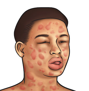
Symptoms can occur immediately after exposure to the antigen and can occur in one or more target organs, such as cardiovascular, respiratory, gastrointestinal, skin, eyes, central nervous system and the urinary system, and other systems. Complaints are often found in the beginning phase is fear, soreness in the mouth, itchy eyes and skin, heat and tingling in the limbs, spasms, hoarseness, nausea, dizziness, weakness and abdominal pain.

In there eyes include conjunctival hyperemia, edema, eye discharge excessive. In allergic rhinitis, allergic shiners can be found, the area below the inferior palpebral dark and swollen. Examination of the nose out in the field there are some signs of allergies, such as: allergic salute, the patients using the palm of the hand rub his nose upward to relieve itching and relieving blockages; allergic crease, transverse skin folds due to the nose; later allergic facies , consisting of mouth breathing, allergic shiners, and teeth abnormalities. The interior of the nasal mucosa examined to assess the color, number, and shape of secretions, edema, nasal polyps, and septal deviation. On the skin are erythema, edema, itching, urticaria, skin feels warm or cold, damp / wet, and diaphoresis.

In the respiratory system occurs hyperventilation, decreased pulmonary blood flow, decreased oxygen saturation, increased pulmonary pressure, respiratory failure, and decreased tidal volume. Upper respiratory tract can be impaired if the tongue or oropharynx involved resulting in stridor. Hoarse voice could not even vote at all if edema continues to deteriorate. Complete airway obstruction is the most frequent cause of death in anaphylaxis. Wheezing breath sounds occur when the lower respiratory tract disrupted as bronchospasm or mucosal edema. In addition, a cough, nasal congestion, and sneezing.

Confused and agitated state followed by a decrease in consciousness to occur coma is a disorder of the central nervous. In the cardiovascular system hypotension, tachycardia, pallor, sweating, signs of myocardial ischemia (angina), endothelial leakage causing edema, accompanied by arrhythmia. While the kidneys, renal hypoperfusion resulting in decreased urine expenditures (oligouri or anuri) due to decreased GFR, which ultimately led to acute renal failure. Besides an increase in BUN and creatinine accompanied by changes in the electrolyte content of the urine.

Hypoperfusion on hepatobilier system, resulting in the central cell necrosis, elevated levels of liver enzymes, and coagulopathy. Symptoms that arise in the gastrointestinal tract as a result of acute intestinal edema and spasm of smooth muscle, such as abdominal pain, nausea, vomiting or diarrhea. Encountered sometimes rectal bleeding caused by intestinal ischemia or infarction.

Bone marrow depression that causes coagulopathy, platelet dysfunction, and DIC can occur in hematological system. While disturbances in neuroendocrine and metabolic systems, there is suppression of the adrenal glands, insulin resistance, thyroid dysfunction, and altered mental status. In a state of shock from aerobic metabolism changes to anaerobic resulting in increased lactic acid and pyruvate. Histologically cracks between cells, the cells swell, mitochondrial dysfunction, and cell leakage.

Hyperthermia related to Dengue Hemorrhagic Fever (DHF)

Dengue Hemorrhagic Fever (DHF) - Hyperthermia Care Plan

NCP DHF - Nursing Diagnosis Interventions : Hyperthermia

Hyperthermia is elevated body temperature due to failed thermoregulation that occurs when a body produces or absorbs more heat than it dissipates. Extreme temperature elevation then becomes a medical emergency requiring immediate treatment to prevent disability or death.

Dengue hemorrhagic fever is a severe, potentially deadly infection spread by certain species of mosquitoes (Aedes aegypti).

Early symptoms of dengue hemorrhagic fever are similar to those of dengue fever, but after several days the patient becomes irritable, restless, and sweaty. These symptoms are followed by a shock -like state.

Bleeding may appear as tiny spots of blood on the skin (petechiae) and larger patches of blood under the skin (ecchymoses). Minor injuries may cause bleeding.

Shock may cause death. If the patient survives, recovery begins after a one-day crisis period.

Early symptoms include: Decreased appetite, Fever, Headache, Joint aches, Malaise, Muscle aches, Vomiting.

Acute phase symptoms include:
  • Restlessness followed by: Ecchymosis, Generalized rash, Petechiae, Worsening of earlier symptoms
  • Shock-like state : Cold, clammy extremities, Sweatiness (diaphoretic)


Nursing Diagnosis and Interventions for Dengue Hemorrhagic Fever (DHF) :

Hyperthermia related to the dengue virus infection.

Goal: Hyperthermia can be resolved

Expected outcomes: body temperature returned to normal

Intervention

1) Observation vital signs, especially temperature.

2) Give cold compress (water) on the forehead and armpits.

3) Change clothes that have been soaked with sweat.

4) Encourage the family to put on clothing that can absorb sweat like cotton.

5) Encourage your family to drink lots of approximately 1500 to 2000 cc per day.

6) collaboration with doctors in Therapy, febrifuge.

Priority Nursing Diagnosis Interventions for Myasthenia Gravis

Myasthenia Gravis Priority Nursing Diagnosis Interventions

Myasthenia gravis (MG) is the most common primary disorder of neuromuscular transmission. Myasthenia Gravis is a type of autoimmune disorder, a neuromuscular disease that usually results to muscle weakness and fatigue primarily in the face. This makes the muscles get tired and weakened more easily than what is normal.

The usual cause is an acquired immunological abnormality, but some cases result from genetic abnormalities at the neuromuscular junction. Much has been learned about the pathophysiology and immunopathology of myasthenia gravis during the past 20 years.

This is often manifested whenever a person suffering from this disorder tries to eat. Within a few minutes of chewing and swallowing, his muscles weaken and get tired, making it hard or even impossible to finish his meal. Often, a few minutes of rest will restore the muscles and the person can continue finishing his meal.

The symptoms of myasthenia gravis include:
  • Droopy eyelids (ptosis)
  • Double vision (diplopia)
  • Difficulty swallowing (dysphagia) with an increased risk of gagging and choking
  • Change in the quality of one's voice
  • Increasing weakness of a particular muscle group during continuous use of those muscles
  • Improved strength of muscles after resting those muscle groups
  • Weak cough
  • Difficulty breathing, leading to respiratory failure (myasthenic crisis)

Nursing Diagnosis and Interventions for Myasthenia Gravis:

1. Impaired gas exchange related to respiratory muscle weakness

Purpose:
Patient will maintain adequate gas exchange:

Interventions:

1. Approach to the client, with communication alternatives, if the client is using a ventilator.
2. Note the oxygenation saturation, with oximetry, especially with activity.
3. Measure respiratory parameters regularly.
4. Collaboration with physicians to anticholinergic medication.
5. Sucktion as required anticholinergic drugs increased bronchial secretion).


2. Self-care deficit related to muscle weakness, general fatigue

purpose:
Patients will be able to do at least 25% of the activities themselves and dress up.

Interventions:
1. Create maintenance schedule to the interval.
2. Give the patient a break in between events.
3. Perform self-care for the patient during a very excessive muscle weakness or include family.
4. Demonstrate energy saving techniques


3. Imbalanced Nutrition Less Than Body Requirements related to dysphagia, intubation, or muscle paralysis.

Purpose:
Caloric intake will be adequate to meet the metabolic needs

Interventions :
1. Assess reflex cough reflex and swallowing disorders before administration by mouth.
2. Stop feeding by mouth if the patient is unable to cope with oral secretions or if the cough reflex swallowing disorders or depressed.
3. Replace the hose and give small meals eaten per-interval if there dysfagia.
4. Record intake and output.
5. Perform nutritional consultation to evaluate calories.
6. Measure the patient's body weight every day.

Fluid Volume Deficit related to Gastroenteritis

Nursng Care Plan for Fluid Volume Deficit related to Gastroenteritis

Fluid Volume Deficit NANDA: The state in which an individual experiences vascular, cellular, or intracellular dehydration

Fluid volume deficit, or hypovolemia, occurs from a loss of body fluid or the shift of fluids into the third space, or from a reduced fluid intake. Common sources for fluid loss are the gastrointestinal (GI) tract, polyuria, and increased perspiration. Fluid volume deficit may be an acute or chronic condition managed in the hospital, outpatient center, or home setting. The therapeutic goal is to treat the underlying disorder and return the extracellular fluid compartment to normal. Treatment consists of restoring fluid volume and correcting any electrolyte imbalances. Early recognition and treatment is paramount to prevent potentially life-threatening hypovolemic shock. Elderly patients are more likely to develop fluid imbalances.

Gastroenteritis is a condition that causes irritation and inflammation of the stomach and intestines (the gastrointestinal tract). An infection may be caused by bacteria or parasites in spoiled food or unclean water. Some foods may irritate your stomach and cause gastroenteritis. Lactose intolerance to dairy products is one example.

Many people who experience the vomiting and diarrhea that develop from these types of infections or irritations think they have "food poisoning," which they may, or call it "stomach flu," although influenza has nothing to do with it.

The incubation period for bacterial gastroenteritis can range from 12 hours to 5 days, depending on the bacteria responsible.

Repeated episodes of diarrhoea are the most common symptom of gastroenteritis. Loose, watery stools are usually passed three or more times within 24 hours. The stools may contain traces of blood and mucus.

Other symptoms of gastroenteritis include:
  • vomiting
  • nausea
  • stomach cramps
  • headaches
  • a high temperature (fever) of 38–39C (100.4–102.2F)

Symptoms of dehydration include:
  • tiredness
  • apathy (a lack of emotion or enthusiasm)
  • dizziness
  • nausea
  • headaches
  • muscle cramps
  • dry mouth
  • pinched face
  • sunken eyes
  • passing little or no urine
  • rapid heartbeat

Nursing Care Plan for Gastroenteritis

Nursing Diagnosis : Fluid Volume Deficit
  • related to
  • nausea
  • excessive loss through feces
  • vomiting and restricted intake

Goal:
  • Fluid requirements will be met with

Outcome criteria there are no signs of dehydration


Intervention and Rational :

1. Monitor intake and output.

2. Give oral fluids and parenteral rehydration in accordance with the program

3. Assess vital signs, signs / symptoms of dehydration and the results of laboratory examination.

4. Collaborative implementation of definitive therapy.

Rational :

1. Provides information to determine the status of fluid balance fluid needs replacement.

2. As an attempt rehydration to replace fluids that come out with feces.

3. Assessing hydration status, electrolyte and acid base balance.

4. Provision of drugs is causally important after the cause of diarrhea in mi